Venous Malformations
Venous Malformations
Overview
Venous malformations refer to congenital and acquired venous diseases involving large-caliber veins, excluding port-wine stains (microvenous malformations). These include cavernous venous hemangiomas, venous tumors, facial venous dilation, limb venous dilation and varicosities, facial sclerosing venous malformations, skin angio-keratoepithelioma, nail bed venous glomus tumors, pyogenic vascular granulomas, dental fibrous hemangiomas, external jugular vein varicosities, and limb honeycomb venous malformations. Except for the latter two, which require surgical treatment, all others can be treated with various laser treatments and flap laser surgery. Laser therapy is divided into laser surgical excision and selective photothermolysis. The former is suitable for pyogenic vascular granulomas and dental fibrous hemangiomas, while the latter is used for cavernous venous malformations, venous lakes, facial venous dilation, limb venous dilation and varicosities, facial sclerosing venous malformations, and skin angio-keratoepithelioma. Deep cavernous venous malformations should primarily be treated with flap laser therapy, with larger and more complex cases requiring a combination of sclerotherapy, partial surgical excision, and radiofrequency thermocoagulation.
Venous malformations mainly affect patients' appearance, cause pain, venous stones, and bleeding, and impair functions such as swallowing, speech, vision, and sexual function. Severe oropharyngeal venous malformations can lead to respiratory and sleep disorders, and laryngeal venous swelling can cause more severe outcomes like suffocation, worsening with age. Systemic multifocal venous malformations often lack effective treatments, impacting quality of life, thus requiring combined clinical treatment methods.
Laser Treatment
1. Continuous Nd: YAG Laser
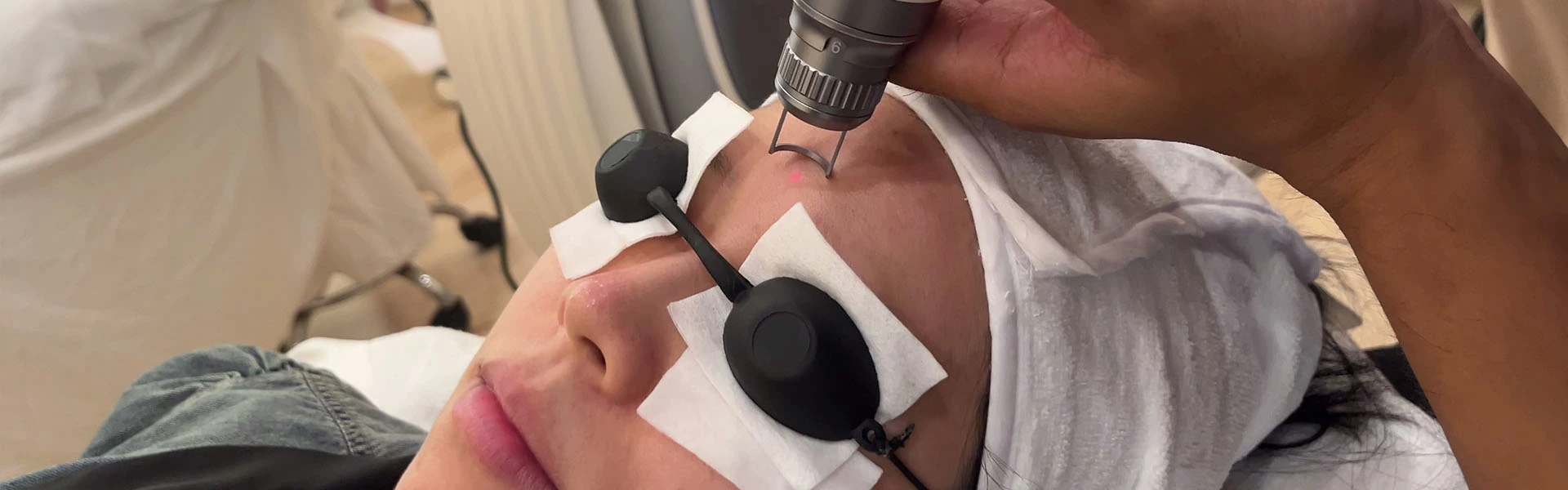
Laser treatment of venous malformations has a long history, with methods established when the Nd: YAG laser was first developed. The infrared laser (1064nm) is strongly absorbed by reduced hemoglobin within the lesion sinuses (65% absorption rate), converting light energy into heat to coagulate, denature, and necrotize lining epithelial cells, inducing fibrosis and closing the lumen to eliminate the mass. Oral and facial superficial varicosities commonly occur in the tongue, gums, soft palate, lips, cheeks, and the posterior molar region, with lesions being solitary or multiple. Another type occurs in those over 60, presenting as isolated lesions on the lips, cheeks, and palate, essentially venous glomus tumors formed due to trauma, often with thrombosis. Effective infrared wavelength lasers include 1064nm Nd: YAG, semiconductor lasers (890nm, 900nm, 980nm), applied continuously at 15-30W, with blue or purple lesions immediately contracting and turning pale. Most superficial venous malformations are cured after 1-2 laser treatments, with larger lesions requiring multiple sessions.
2. Long-Pulse Nd: YAG Laser
Venous malformations involving facial skin and mucosa are best treated with long-pulse Nd: YAG lasers, such as Candela’s Gente YAG laser and Cynosure’s Cynergy laser, offering excellent treatment and cosmetic results for facial and body venous malformations. Treatment parameters vary based on lesion size, with Candela’s laser using 180-240J/cm² dynamic cooling set to 20-30ms/20ms/20ms, and Cynosure’s using 20-40J/cm², with a 20-30ms pulse width and 1-2 level cold air cooling. Incorrect parameter settings can cause adverse reactions or severe complications.
3. 980nm Semiconductor Laser
Since 2000, semiconductor lasers have gained popularity, using wavelengths of 532nm, 810nm, and 980nm. The 980nm laser, chosen for its deeper penetration, is used for photocoagulation of venous malformations. Local infiltration or block anesthesia with 2% lidocaine is used. The laser operates in continuous mode at 13-16W, maintaining a non-contact distance of 0.5-1.0cm from the lesion until effective contraction and pallor occur. Over-treatment can cause intraoperative bleeding and lesion rupture. Postoperative care includes prophylactic antibiotics and anti-inflammatory medications to reduce pain and other complications.
4. Carbon Dioxide Laser
The carbon dioxide laser, using a light knife mode, is absorbed strongly by water molecules at a 1600nm infrared wavelength, with shallow penetration. It is suited for lesions with thrombosis. After local anesthesia, a 5W power setting is used for initial incision design, followed by 10-20W focused mode cutting to expose and excise the lesion, with layered suturing to close the wound.
5. Flap Laser Treatment
Deep venous malformations covered by normal tissue require flap laser treatment, as direct laser exposure cannot penetrate to the lesion. Traditional methods include surgery, sclerotherapy, and copper needle insertion but have limitations and recurrence risks. Recently, a microwave thermocoagulation combined with surgical excision method has shown advantages in reducing bleeding, increasing excision rates, and improving outcomes. Nd: YAG laser irradiation experiments on animal facial nerves established safe laser dosage parameters, preventing nerve damage during clinical application. Flap laser treatment involves sequential dissection and laser coagulation of the lesion at 70-100J/cm², with ice saline irrigation to prevent thermal nerve damage. This method effectively reduces intraoperative bleeding, simplifies procedures, and protects facial nerves, improving outcomes for deep venous malformations in various regions, including the parotid, masseter, submandibular, and pharyngeal areas, with significant symptom relief.
Reports from 1999 to 2005 detail successful flap laser treatment of 362 deep venous malformation cases, with a high lesion elimination rate and minimal complications. Semiconductor 980nm laser has also been widely adopted across surgical disciplines, preserving normal tissue and structure. Intraoperative hemostasis with infrared laser greatly improves surgical safety and efficiency, reducing anesthesia dosage and surgical risks.
When treating limb deep venous malformations, a tourniquet is essential to minimize bleeding and maintain a clear surgical field. Preoperative assessment of lesion size and volume is crucial, avoiding excessive single-session treatment. Different venous types require specific laser techniques, with honeycomb venous malformations contraindicated due to bleeding risk. MRI imaging aids in differentiation and treatment planning.
Flap laser treatment is suitable for 80% of deep venous malformations, with combined methods for large lesions ensuring effective control and normal tissue function preservation. Ongoing research focuses on new treatment methods.
Main Complications of Laser Treatment for Venous Malformations
- Postoperative Swelling: Occurs 24-72 hours post-treatment, requiring prophylactic medication like steroids and antibiotics.
- Lesion Ulceration and Pain: Common 1-2 weeks post-laser, preventable by monitoring laser dosage and immediate tissue response.
- Postoperative Bleeding: Occurs within two weeks, preventable by avoiding excessive laser dosage. Elderly and frail patients need enhanced postoperative nutrition and limited treatment site movement.
Flap laser treatment’s main complication is nerve damage, preventable by careful surgical exposure, hemostasis, and ice saline cooling during laser use. Other complications include excessive exudation and dead space formation, managed by tight compression bandaging and appropriate drainage device use.
Source: Venous Malformations
 Ciellulu Laser - Facial Machine Supplier