Telangiectasia

Telangiectasia
Telangiectasia refers to superficial skin blood vessels that are visible to the naked eye. 10% to 15% of adults and children have noticeable facial telangiectasia.
I. Causes and Pathogenesis
All types of telangiectasia are related to the release of vasoactive substances. Triggers include chronic photo damage, alcohol, hypoxia, estrogen, and corticosteroid hormones (topical or systemic), chemicals, various bacterial and viral infections, multiple physical factors, ultimately leading to the neovascularization of capillaries and venules. Damage and pressure from surgical excision, cosmetic surgery, or rhinoplasty can also promote new blood vessel formation, leading to telangiectasia. Some families exhibit autosomal dominant inheritance.
II. Clinical Manifestations
Facial telangiectasia has no age or gender differences. The diameter of dilated capillaries generally ranges from 0.1 to 1.0 mm, consisting of dilated venules, capillaries, or small arteries. Telangiectasia originating from small arteries and capillaries has a smaller diameter, is bright red, and usually does not protrude above the skin surface. Telangiectasia originating from venules is larger, blue, and often protrudes above the surface. Capillary loop telangiectasia initially appears as small, red spots that gradually enlarge, turning purple or blue as venous pressure and venous return increase. Based on clinical manifestations, telangiectasia is classified into four types: simple or linear, branching, spider angioma, and nodular.
Red linear and branching types are more common on the face and lower limbs, especially the nose, central cheeks, and chin. Nodular type is often a skin manifestation of hereditary diseases such as Osler-Weber-Rendu disease and can also be seen in collagen vascular diseases. One of the typical skin manifestations of rosacea is telangiectasia, with deeper vascular dilation and increased fine blood vessels causing facial erythema and flushing. Civatte's poikiloderma is a clinical symptom caused by chronic excessive sun exposure, presenting as reticulate brown pigmentation, scattered and fused telangiectasia, and prominent telangiectasia on the lower face, neck, and anterior chest.
III. Diagnosis and Differential Diagnosis
Diagnosis can be made based on facial telangiectasia that blanches under pressure. This disease mainly needs to be differentiated from the following diseases:
- Telangiectatic Lupus Erythematosus: A rare type of lupus erythematosus, possibly related to photosensitivity and autoimmune-induced vascular changes.
- Persistent Pigmented Purpuric Dermatosis: A special type of pigmented urticaria.
- Idiopathic Telangiectatic Purpura: Cause unknown, commonly seen on the lower legs of adults, resulting from lymphocytic perivasculitis, presenting as yellow-red ring-shaped, spotty petechiae and telangiectasia, which may last for years.
- Alcoholic Hemorrhagic Telangiectasia: Autosomal dominant inheritance, common after puberty, with lesions on the back of the hands, face, scrotum, often with spider angioma-like lesions around them. Common on the lips, tongue, nasal mucosa, cheeks, or gums, characterized by bleeding at the lesion site.
- Ataxia Telangiectasia: Autosomal recessive inheritance, with onset at 2-3 years, characterized by cerebellar ataxia and telangiectasia of the eyes and skin. Initially affecting the bulbar conjunctiva, then spreading to the eyelids, cheeks, ears, neck, and elbows, often accompanied by nystagmus, café-au-lait spots, white hair, and premature aging.
- Congenital Marble Skin Telangiectasia: Generalized reticulate cyanosis at birth, with spider angiomas and vascular keratomas that may resolve.
- Spider Angioma: Can be congenital or acquired, with the former common in children and the latter in liver disease and pregnant women.
- Generalized Essential Telangiectasia: Onset in childhood or adolescence, initially affecting the lower legs and gradually spreading to the thighs, abdomen, and upper arms, with widespread telangiectasia but no systemic disease. Skin may become atrophic, thin, loose, and less elastic, causing telangiectasia, especially on the face and lower limbs.
IV. Treatment
-
General Treatment:
First, identify and address the underlying cause and trigger factors. Treat the primary disease while addressing telangiectasia. -
Laser Treatment:
- Laser Treatment Devices:
- Carbon Dioxide Laser:
Uses the absorption of carbon dioxide laser by tissue water for treatment. However, due to its non-selectivity, it removes normal skin along with the affected blood vessels, making it unsuitable for clinical use. - Argon Laser:
Laser output power ranges from 0.8 to 2.9W, exposure times of 50ms, 0.2s, and 0.3s, with a spot diameter of 0.1mm and 1mm. Effective but with notable side effects, including pitted scars, hypopigmentation, hyperpigmentation, and lesion recurrence. - Copper Vapor/Copper Bromide Laser:
Wavelengths of 578nm (yellow) and 511nm (green), considered quasi-continuous lasers. Comprising a series of pulses (15,000 sub-pulses per second), these lasers' tissue effects are similar to continuous lasers but can also be divided into 20-50ms pulses or used with a scanning device. With a pulse width of 20-50ms, they are safely used for telangiectasia treatment, offering better efficacy than argon lasers and comparable to pulsed dye lasers. A fine scab may form post-treatment. - Pulsed Dye Laser:
Improvement rate exceeds 50% after 1-2 treatments. Over 97.5% of patients achieve good results with multiple treatments. Larger vessel diameters require longer pulse widths and more treatments, with treated areas turning blue-purple, fading in 7-14 days without scarring. - Long-Pulse Nd:yag Laser:
For larger, deeper vessels, deeper penetration lasers like Ndare needed. Absorption by hemoglobin is only 1/10th that of pulsed dye lasers, requiring over ten times the energy density for similar effects, increasing scar risk. Care must be taken to protect the epidermis and lower skin temperature to prevent burns. - Dual-Wavelength Laser:
Effective for telangiectasia treatment below the purpura threshold, reducing scar risk. - Intense Pulsed Light (IPL):
Effective for large-area facial telangiectasia, especially for facial flushing and fine capillaries. For significant telangiectasia and larger vessels, pulsed dye laser and Ndlaser are better choices.
- Carbon Dioxide Laser:
- Post-Laser Skin Care:
Specific post-laser skin care methods can be found in the treatment for port-wine stains.
- Laser Treatment Devices:
V. Additional Resources
Telangiectasia and Autoimmune Disease
Source: Telangiectasia
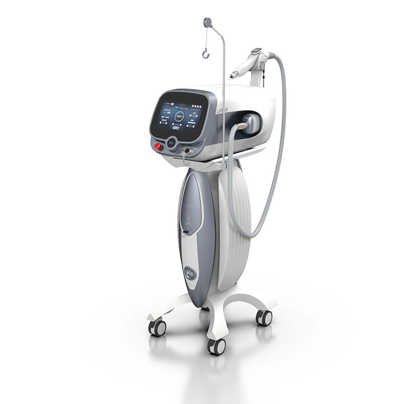
 Ciellulu Laser - Facial Machine Supplier