Spider Telangiectasia

Spider Telangiectasia
Spider telangiectasia, also known as spider nevus, is an acquired benign vascular disease with an incidence of 10% to 15% in healthy adults and children. Most cases are unrelated to dermatological or internal diseases. Many women develop spider telangiectasia during pregnancy or while taking oral contraceptives, with spontaneous resolution 6 to 9 months postpartum or after discontinuing the medication. Patients with liver diseases often have multiple and prominent spider nevi. The rash of a spider nevus consists of a central arteriole with radially distributed small blood vessels, resembling a spider’s body and legs.
I. Causes and Pathogenesis
Spider nevi are not due to vascular proliferation but to the dilation of existing vessels. Liver cirrhosis, liver cancer, or other liver diseases can cause rapid onset and multiple, prominent spider nevi. High estrogen levels are often detected in the blood. If spider nevi are accompanied by palmar erythema, pale nails with congested nail beds, liver cirrhosis should be considered, often associated with splenomegaly, ascites, jaundice, and tremors. Children with liver disease frequently have multiple spider nevi, although healthy children can also have more than five spider nevi.
II. Clinical Manifestations
This condition is common in preschool and school-age children, with a peak incidence at ages 7 to 10. By age 15, about 40% of girls and 32% of boys will have at least one rash, which can persist for years. Rashes commonly occur on the face, neck, upper torso, and arms, with children's hands and fingers frequently affected. Spider nevi are typically bright red, with a central small red papule (diameter less than 1mm) surrounded by radiating small blood vessels. The entire rash diameter is 0.5 to 1 cm. Pressing on the rash makes it disappear; releasing the pressure shows blood refilling from the central arteriole to the surrounding small vessels. Sometimes, the central arteriole's pulsation can be felt. Common sites include the face, beneath the eyelids, or the zygomatic area, as well as the hands, forearms, and ears. Pregnant women and liver disease patients may also have palmar erythema. Severe internal diseases often result in multiple spider nevi.
III. Pathological Features
The central lesion consists of an ascending artery with smooth muscle in its wall, or vascular glomerulus cells between endothelial cells and the internal elastic membrane. The artery expands into a thin-walled ampulla beneath the epidermis, with slender arterial branches radiating outward, dividing into many capillaries.
IV. Diagnosis and Differential Diagnosis
Diagnosis is based on the typical appearance of a central arteriole with radially distributed small vessels. Pressing the central arteriole causes it to blanch; releasing the pressure quickly refills the radial vessels with blood. Differential diagnosis includes telangiectasia diseases, where lesions are clusters of small dilated capillaries, appearing purplish-red or bright red, with no pulsation.
V. Treatment
- General Treatment
Most children's spider nevi resolve spontaneously without special treatment, although complete resolution may take years. For pregnant women, rashes often resolve 6 to 9 months postpartum or after stopping oral contraceptives. Spider nevi in liver disease patients correlate with liver function changes. - Laser Treatment
- Laser Treatment Devices
- Pulsed Dye Laser:
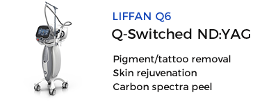
Highly effective with a 70% cure rate after one treatment, 2 to 3 treatments result in near-complete resolution. Initial treatment targets the center of the spider nevus with 1-2 pulses at 6.5-7.7J/cm². For lesions larger than 5mm, treat the radiating vessels with overlapping 10% pulses. Effective for adult spider nevi as well, with side effects including purpura and temporary pigment changes. - 532nm Frequency-Doubled Nd:YAG:
Effective for spider nevi, typically using a 3-4mm spot diameter, 10ms pulse width, and 12-14J/cm² energy density. - Long-Pulse NdLaser:
Initially targets the central papule with 1-2 pulses. Radial vessels often disappear with this treatment. If not, treat along the vessel's path. - Dual-Wavelength Laser:
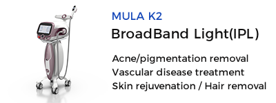
Safe and effective for spider nevi treatment. - Intense Pulsed Light (IPL):
Very effective for spider nevi. Prior to treatment, match the lesion area and shape with a template on a whiteboard. Use a double pulse with a 2.5-3.0ms pulse width and 25-40ms delay. Significant improvement is usually seen after one session.
- Pulsed Dye Laser:
- Post-Laser Skin Care:
Follow specific post-laser skin care methods as outlined for port-wine stains.
- Laser Treatment Devices
Pyogenic Granuloma
Pyogenic granuloma (granuloma pyogenicum) is an acquired vascular tumor, distinct from inflammatory granuloma. The name "pyogenic granuloma" is a misnomer since the disease is neither infectious nor granulomatous.
I. Causes and Pathogenesis
The exact cause and pathogenesis of pyogenic granuloma are unclear. Historically, trauma was considered a primary cause, but large studies show only 7% of lesions follow trauma. Potential factors include trauma, endocrine changes, viral oncogenes, minor arteriovenous malformations, abnormal vascular endothelial growth factor, and cytogenetic abnormalities. Pyogenic granulomas often develop rapidly at recently injured sites, possibly representing an abnormal response of vascular or fibrous tissue to injury.
II. Clinical Manifestations
Pyogenic granuloma can occur at any age and shows no significant gender difference. It commonly occurs in trauma-prone areas such as the face, head, fingers, feet, and upper torso. Lesions are typically single, bright red, fragile, polygonal papules or nodules with a stalk, ranging from a few millimeters to several centimeters. They are soft, bleed easily, and may ulcerate, forming a granuloma-like surface covered with a brown-black scab. Lesions appear suddenly and grow rapidly, eventually shrinking and becoming fibrotic if untreated.
Pyogenic granulomas can be segmentally distributed, subcutaneous, intravenous, or drug-induced (e.g., by retinoids or chemotherapy). Granulomas during pregnancy often occur in the mouth, especially the gums.
III. Pathological Features
Lesions exhibit inward growth of normal epidermal tissue, forming a constriction band resembling a collar. Endothelial cells proliferate in a lobular pattern. In mature lesions, endothelial cells cluster into solid masses, with most areas showing lumen formation, varying from slit-like to significantly dilated. Endothelial cells in many lumens are swollen and protrude into the lumen. Early lesions show minimal inflammation, while older lesions often have secondary inflammatory changes.
IV. Diagnosis and Differential Diagnosis
Diagnosis is based on the occurrence at any age and any site, often following trauma. If diagnosis is challenging, a biopsy can confirm it. Differential diagnosis primarily includes strawberry hemangioma, characterized by a soft, dark red or bright red nodular appearance that appears within weeks after birth, grows rapidly in a few months, and regresses gradually after 1-2 years.
V. Treatment
- General Treatment
If there are clear traumatic triggers, remove the cause. Pregnancy-related granulomas often resolve spontaneously postpartum, so treatment is usually deferred until after delivery. - Drug Treatment
Topical imiquimod and retinoid creams can treat pyogenic granuloma. Recurrent lesions with satellite lesions may require local or systemic corticosteroid injections. Large lesions may be treated with intralesional bleomycin injections. - Surgical Treatment
Surgical options include complete excision, curettage, and electrodessication of the base to reduce recurrence risk. Sclerotherapy, chemical cauterization (e.g., silver nitrate, trichloroacetic acid), and cryotherapy are effective. - Laser Treatment
- Laser Treatment Devices:
- Argon and Carbon Dioxide Lasers:
Effective for pyogenic granuloma, with treatment endpoints being lesion turning gray-white. Repeat treatment every 3-4 weeks if needed. - Pulsed Dye Laser:
Treatment efficacy is unpredictable. Thick lesions may resist penetration, requiring glass plate compression to reach deeper vessels before removing the plate to treat superficial lesions. Overlap pulses in place until the color turns gray-white, similar to continuous lasers, copper vapor lasers, and 577nm dye lasers. - Dual-Wavelength Laser:
Shows significant efficacy in treating pyogenic granuloma, enhancing specific laser absorption and penetration depth, typically requiring 1-3 treatments.
- Argon and Carbon Dioxide Lasers:
- Post-Laser Care:
Follow specific post-laser skin care methods as outlined for port-wine stains.
- Laser Treatment Devices:
Source: Spider Telangiectasia
 Ciellulu Laser - Facial Machine Supplier