Port Wine Stain
Port Wine Stain
Port Wine Stain (PWS)
Port Wine Stain (PWS) , also known as nevus flammeus or capillary malformation, is a relatively flat and rarely raised plaque composed of dilated capillaries. It is a congenital capillary malformation.
Etiology and Pathogenesis
The etiology and pathogenesis of PWS are unknown. It is generally believed to result from errors in gene expression, translation, or protein synthesis during embryonic development, with genetic factors involved. It usually appears at birth or shortly after and grows in proportion to the body, never regressing. The incidence is 0.3% to 0.5% in newborns. PWS is usually sporadic but familial cases have been reported, with autosomal dominant inheritance. Some patients develop PWS later in life, possibly triggered by trauma, oral contraceptives, or prolonged sun exposure. The capillaries in the lesions are dilated but have normal endothelial cells, and immunohistochemical staining cannot distinguish them from normal dermal capillaries. However, S100 protein staining shows a significant reduction in sympathetic nerve endings in the superficial dermal vessels, which may relate to changes in vascular tension and capillary dilation.
Clinical Manifestations
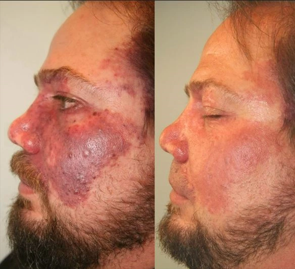
PWS commonly occurs in infants or children, with similar rates in males and females. Lesions often appear on the face, neck, and scalp, usually unilateral but sometimes bilateral, occasionally involving mucous membranes. Most PWS are very superficial, with an average depth of 0.46mm. New lesions present as well-defined red patches, which can appear anywhere on the body but 90% occur on the head and neck, especially in the first and second branches of the trigeminal nerve. Initially, the lesions are pink and gradually turn deep red or purple with age. The color change is unrelated to the depth or thickness of the vessels. Over time, the vessels become tortuous and dilated, thickening the lesions and giving a cobblestone appearance, sometimes forming nodules. Studies show that by age 46, about two-thirds of patients develop skin thickening and nodule formation, typically starting at around 37 years old. Nodules can easily ulcerate and bleed. Even in infancy, PWS can develop pyogenic granulomas. Lesions generally persist for life and rarely regress spontaneously. PWS can also cause hypertrophy of the underlying skin, soft tissues, and bones.
In addition to cosmetic concerns, PWS can be associated with glaucoma (with a 45% chance if it involves the trigeminal nerve's upper branch), ulceration, bleeding, and secondary infections. About 9.5% of facial PWS patients have eye or nervous system abnormalities, with larger lesions more likely to have systemic complications.
Under in vivo videomicroscopy, PWS shows two types: (1) tortuous, superficial, and dilated vascular loops (spot type); (2) superficial, parallel, and dilated vascular plexus (ring type). Some lesions have characteristics of both types. The first type responds better to pulsed dye laser treatment, while the second type is deeper and has connecting vessels between dilated and parallel vascular plexus.
Pathological Features
Microscopically, PWS lesions show clustered dilated capillaries and mature endothelial cells in the upper and mid dermis. Capillary dilation increases with age and can extend into the deep dermis and subcutaneous tissue without endothelial cell proliferation. Surrounding the vessels are loosely arranged collagen fibers, and the lumen is filled with red blood cells. In infancy, there are no significant abnormalities; in adulthood, only subpapillary vascular dilation is seen.
Diagnosis and Differential Diagnosis
Diagnosis is based on the presence of red or dark red patches at birth or appearing shortly after, enlarging with body growth and not regressing spontaneously.
Differential diagnosis includes:
- Early Congenital Hemangioma: Diagnosis requires observing lesion progression, which is usually rapid in hemangiomas.
- Infantile Port Wine Stain: Also known as simple nevus, nevus flammeus, or median capillary malformation, often involving the forehead, nose, upper lip, occipital scalp, or eyelids. Usually pale pink, it typically fades or disappears by 1-2 years of age, except for lesions on the midline forehead or sacral area.
Treatment
- Surgical Treatment: For larger hemangiomas or visceral vascular tumors.
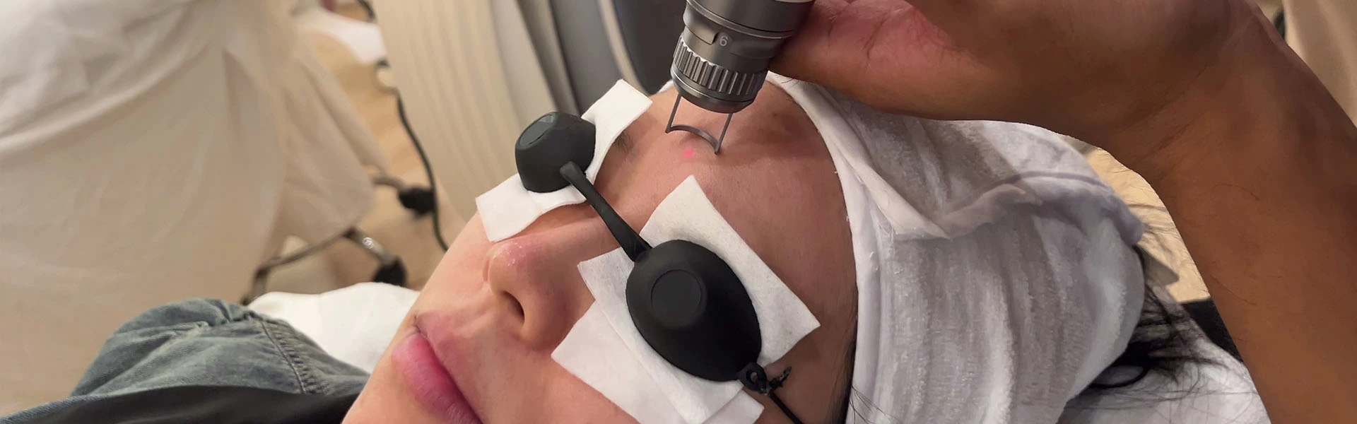
- Laser Treatment:
- Pulsed Dye Laser (PDL): The gold standard for PWS treatment, with wavelengths of 577nm, 585nm, or 595nm, selectively absorbed by oxyhemoglobin with minimal damage to other epidermal components. Despite the optimal absorption peak for oxyhemoglobin being 420nm, this wavelength penetrates too deeply to reach dermal vessels. Compared to 585nm, 595nm has less absorption specificity but penetrates deeper, making it suitable for deeper vessels. Pulse durations typically range from 0.45 to 40ms, with fine vessels in children best treated with a 0.5ms pulse duration and larger vessels with longer pulses. Energy density is usually 5-10J/cm2. To alleviate pain, topical anesthetics can be applied before treatment. Treatment endpoints are darker lesion color and purpura, with common side effects including blisters, post-inflammatory hyperpigmentation, hypopigmentation, skin sensitivity, and scarring. PDL is particularly effective for small capillaries in children but less effective for proliferative or nodular lesions. Studies show an average improvement rate of 75% after 2.5 treatments. Generally, 5-10 treatments achieve clearance or improvement, with superficial lesions responding faster, achieving 95% improvement after 1-2 treatments. Adults respond less favorably, with about 20% resistant to treatment and a first-treatment improvement rate of about 50%. Early treatment benefits include faster improvement, fewer treatment sessions, smaller lesion area, and lower anesthesia requirements.
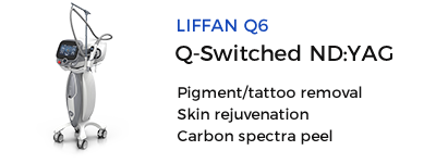
- NdLaser: Effective for resistant or hypertrophic PWS with deeper penetration and longer pulse durations (1064nm). While similar in absorption to 585nm lasers, the 1064nm laser has a lower absolute absorption and scattering coefficient. PWS lesions are usually 3-5mm below the dermis, beyond the reach of PDL, making longer wavelengths viable. Long-pulsed Ndlasers rarely cause purpura but can lead to blistering and scarring. Typical pulse durations are 10-50ms, with energy densities of 40-120J/cm2, treatment intervals of 1-2 months, and 4-10 sessions needed.
- Dual-Wavelength Laser: Combining PDL and long-pulsed Ndlasers, where PDL's wavelength closely matches the oxyhemoglobin absorption peak but has limited efficacy for deep, large vessels due to its shorter wavelength and superficial penetration. Dual-wavelength lasers use a sequential emission mode, first emitting PDL to convert oxyhemoglobin to methemoglobin, followed by Ndlaser, significantly improving treatment specificity and efficacy while reducing purpura and scarring. Typical combinations are PDL pulse durations of 0.25-10ms, energy densities of 4-9J/cm2; 1064nm laser pulse durations of 15-40ms, energy densities of 20-60J/cm2, with output intervals of 500-1000ms and treatment intervals of 6-8 weeks, adjusted based on lesion characteristics and treatment response. Continuous air cooling and topical anesthetics (e.g., 2% lidocaine + 2% prilocaine cream) are needed. Adverse reactions include pain, erythema, edema, blistering, hypopigmentation, and scarring.
- Frequency-Doubled NdLaser: An option for recurrent PWS, with a shorter wavelength (532nm) limiting penetration depth and causing more side effects, such as transient pigmentation changes and scarring, due to high melanin absorption. It's recommended for refractory PWS with appropriate pulse durations to avoid purpura.
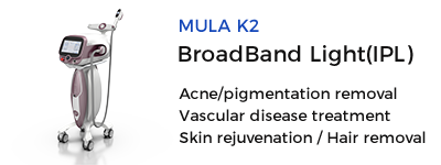
- Intense Pulsed Light (IPL): A broad-spectrum pulsed light with good efficacy, especially for PDL-resistant lesions. Parameters for LumenisOne system include single-pulse treatment with 560nm/590nm/640nm filters, pulse durations of 4-10ms, energy densities of 12-22J/cm2, double-pulse treatment with 560nm/590nm filters, pulse durations of 3.5-4.0ms, energy densities of 17-30J/cm2, and pulse intervals of 20-33ms, with treatments every 20-40ms.
Post-Laser Skin Care After laser treatment, the skin barrier is compromised, necessitating proper post-treatment skin care and appropriate medical skincare products to repair the damaged skin.
- Reducing Erythema and Exudation: Depending on immediate post-treatment skin response, use masks containing anti-inflammatory, moisturizing ingredients or cold compresses with ice packs wrapped in gauze. If the skin turns white post-treatment, cold compresses for about 30 minutes are recommended; for normal congestion and redness, 15 minutes is sufficient, avoiding friction. For significant erythema, swelling, or bleeding, use 3% boric acid solution or 5% furacilin solution cold compresses.
- Reducing Inflammatory Response and Preventing Infection: To prevent postoperative wound infection, apply mupirocin ointment, erythromycin ointment, or gentamicin injections topically. For extensive treatment areas and severe inflammation, oral prednisone 10mg three times daily for three days can enhance anti-inflammatory effects.
- Promoting Wound Healing: Basic fibroblast growth factor (BFGF) is an important mitogen promoting wound healing, tissue repair, and regeneration. Post-laser treatment, apply BFGF gel or spray evenly on the wound to promote healing.
- Promoting Skin Regeneration and Repair: Rebuilding normal skin structure and restoring physiological functions are crucial due to varying degrees of damage to the skin's lipid barrier, stratum corneum, aquaporin, brick wall structure, and basal layer. Using suitable medical skincare products with anti-inflammatory and moisturizing effects for 3-6 months post-treatment is essential.
- Sun Protection: Wear a sun hat, long-sleeve cotton clothing, and use an umbrella (preferably UV-protective) when outdoors. Apply sunscreen with a high SPF (above 30) and UVA protection (PFA ++ or higher). Avoid prolonged sun exposure between 10 am and 4 pm. High-safety, effective sunscreens are needed to prevent post-laser pigmentation changes.
Other Treatments
- Radiation Therapy: Using shallow X-ray irradiation effectively treats capillary hemangiomas.
- Cryotherapy: Liquid nitrogen cryotherapy, tailored to the size and shape of lesions, is mainly used for capillary hemangiomas, but it can cause scarring.
- Photodynamic Therapy: An emerging treatment option for PWS, potentially becoming the future direction. It involves injecting a photosensitizer into the blood, forming a high concentration in the vessels, and then using a specific wavelength of laser light to produce reactive oxygen species, causing endothelial cell damage and vessel wall destruction while sparing the surrounding tissues. Photosensitizers like hematoporphyrin derivatives (HpD) or hematoporphyrin monomethyl ether (HMME) are used after a skin test. HpD or HMME is intravenously injected at 4.5-5.5mg/kg, followed by laser irradiation. Treatment intervals are usually around one month, with HpD-PDT requiring one-month light avoidance and HMME-PDT needing 1-2 weeks. Ideal for pink lesions in children, less effective for thick, purple lesions, and carries a risk of scarring with prolonged laser exposure. HMME-PDT has milder postoperative reactions, shorter healing periods, greater safety, and shorter light avoidance periods compared to HpD-PDT.
Additional Resources
Hematoporphyrin Monomethyl Ether Photodynamic Therapy of Port Wine Stain: Narrative Review[NCBI]
Source: Port Wine Stain

Port Wine Stains

 Ciellulu Laser - Facial Machine Supplier