Cherry Angioma

Cherry Angioma
Cherry Angioma (cherry angioma), also known as senile angioma, is a small localized red spot or papule composed of dilated blood vessels.
I. Causes and Pathogenesis
Currently, little is known about the cause of cherry angioma. Cherry angioma is formed by the proliferation of dilated venules.
II. Clinical Manifestations
This disease is common in the elderly. It can occur on any part of the body, especially on the abdomen. The shape of cherry angiomas varies. Initially, they are small red spots that can develop into larger raised papules or polygonal papules. The typical rash is red, but it can also appear purplish-red. When thrombi block the vascular cavity, the rash turns dark brown or almost black, which may be mistaken for malignant melanoma.
III. Pathological Features
In the early stages, many newly formed capillaries with narrow lumens and lobules mainly formed by endothelial cells can be seen in the sub-papillary layer. Subsequently, the capillaries gradually dilate, and many moderately dilated capillaries lined with flat endothelial cells can be seen. Edema of the stroma, homogenization of collagen fibers, and slight atrophy or disappearance of the epidermis can occur, often surrounding most of the angioma like a collar.
IV. Diagnosis and Differential Diagnosis
Diagnosis can be made based on the bright red or cherry-colored papules that occur on the trunk of the elderly. If it is difficult to confirm the diagnosis based on clinical features alone, a biopsy can be performed for pathological examination. This disease mainly needs to be differentiated from the following diseases:
- (a) Spider Nevus (spider nevus): When larger, it resembles smaller senile angiomas, but the lesions of this disease do not have dilated capillaries around them.
- (b) Pyogenic Granuloma (pyogenic granuloma) and Bacillary Angiomatosis (bacillary angiomatosis): Pyogenic granuloma shows significant endothelial cell proliferation, while cherry angioma does not. Bacillary angiomatosis shows granuloma fragments and epithelioid endothelial cells at the lesion site.
V. Treatment
- (a) Physical Therapy Physical therapy for cherry angiomas includes chemical peeling, electrocautery, and curettage.
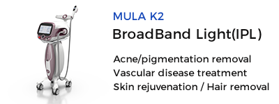
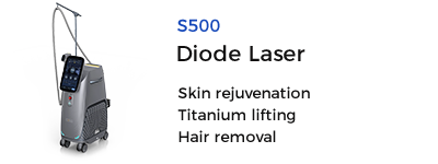
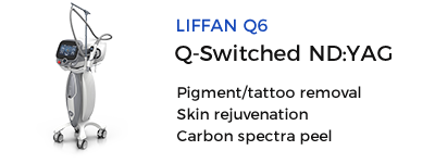
- (b) Laser Therapy Laser options include ultrapulse carbon dioxide laser (for smaller lesions), laser, 532nm KTP laser, Nd:yag laser, IPL, dual-wavelength laser, etc. After treatment with KTP laser and Ndlaser, the surface of the rash may scab, and purpura may form with laser treatment.
- (c) Post-Laser Care For specific methods of post-laser skin care, refer to the care for port-wine stains.
Source: Cherry Angioma
 Ciellulu Laser - Facial Machine Supplier