Angiokeratoma

Angiokeratoma
Angiokeratoma, also known as angio-keratosis, is a skin disease characterized by capillary dilation in the upper dermis and hyperkeratosis of the epidermis.
I. Causes and Pathogenesis
The causes and pathogenesis of angiokeratoma are unclear. Possible factors include genetic predisposition, pregnancy, trauma, subcutaneous hematoma, and tissue hypoxia.
II. Clinical Manifestations
-
Acral Angiokeratoma
Acral angiokeratoma, also known as Mibelli angiokeratoma, is autosomally dominant and common in children or adolescents, with a higher incidence in females. Often preceded by a history of frostbite or chilblains, there are reports of multiple family members being affected simultaneously. It commonly occurs on the dorsal sides of fingers and toes, knees, elbows, occasionally finger joints, palms, soles, and ears. Typically symmetrical. Lesions come in two forms: pinhead to millet-sized macules or papules, several to dozens, rough, keratotic, purple or dark purple, sometimes blanching upon pressure; or nodular lesions, 2-8mm in diameter, purplish-red or gray-brown, hyperkeratotic or warty, with dilated capillaries or blood scabs centrally, bleeding easily after trauma. No significant subjective symptoms. -
Scrotal Angiokeratoma
Also known as Fordyce angiokeratoma, occurring mainly in middle-aged and elderly men, occasionally in the labia, increasing with age. Multiple dome-shaped papules on the scrotum, 1-4mm in diameter, initially bright red and soft, later dark red or purple and hard with mild warty hyperplasia. Often aligned along superficial veins or scrotal skin lines, smooth surface, sometimes slightly scaly, blanching upon pressure. Occasionally found on the penis or glans, rarely on the lower legs, thighs, and conjunctiva. Generally asymptomatic, sometimes mildly itchy, bleeding easily after injury. Often associated with epididymal tumors, hernia, varicocele, oral mucosal varices, and scrotal elastin deficiency. -
Solitary Angiokeratoma
Common in young people, typically single but occasionally multiple papules or nodules, 2-10mm in diameter, initially bright red and soft, later turning blue or black with hyperkeratosis and increased hardness. Common on the lower limbs, asymptomatic, sometimes mistaken for malignant melanoma. -
Localized Angiokeratoma
Usually present at birth or in childhood/adolescence, rare. Common on the lower legs and feet, occasionally on the back and forearms. Initially single, occasionally multiple, light purple-red clustered papules or cystic nodules filled with blood, later merging into irregular or linear plaques, warty surface. Typically enlarges with age and develops new lesions, usually a few centimeters in size, occasionally larger, continuously expanding. Clinically, localized angiokeratoma resembles localized lymphangioma, with some superficial nodules containing blood and others lymph fluid, termed intermediate type. This type can coexist with scrotal angiokeratoma, oral varices, and hypertrophic port-wine stains or cavernous hemangiomas. -
Diffuse Trunk Angiokeratoma
Caused by a deficiency of α-galactosidase, this rare genetic disease is X-linked recessive, common in children and adolescents, primarily involving lipid deposition in small skin and visceral blood vessels, characterized by diffuse angiokeratoma-like lesions with cardiovascular and renal damage.
III. Pathological Features
This disease causes hyperkeratosis or irregular acanthosis, papillomatous hyperplasia, and marked capillary dilation in the upper dermis, with red blood cells and thrombi in the lumens. The deep dermis and subcutaneous tissue also show vascular dilation, congestion, endothelial cell proliferation, and sometimes cavernous hemangiomas. Mixed lymphatic and blood-filled lumens may be seen.
IV. Diagnosis and Differential Diagnosis
Diagnosis is usually straightforward based on history, clinical presentation, and pathological examination. The main differential diagnosis is malignant melanoma: angiokeratoma is benign but can resemble melanoma in appearance, requiring excision and pathological confirmation.
V. Treatment
-
Surgical Treatment
For a few lesions, surgical excision is an option; for multiple lesions, electrocautery or cryotherapy can be used. -
Laser Treatment
- Laser Devices:
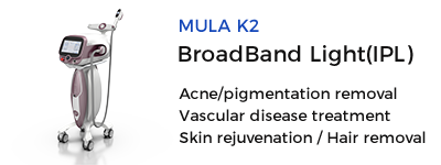
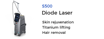
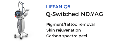
- For proliferative, non-hyperkeratotic lesions, treatments include copper vapor laser, frequency-doubled 532KPT laser, Ndlaser, pulsed dye laser, dual-wavelength laser, and intense pulsed light.
- For warty, hyperkeratotic lesions, ultrapulse carbon dioxide laser is effective. High-energy ultrapulse CO2 laser ablation is a simple and effective method. After routine disinfection and local anesthesia, the lesion is scanned with a power of 5-10W, simultaneously burning and vaporizing the surface while coagulating the underlying blood vessels. Post-burn, the charred tissue is wiped off to reveal uniform yellow-white dermal tissue, indicating successful angiokeratoma removal. If bleeding occurs after wiping off the char, lower power density laser coagulation is used until hemostasis. For scrotal angiokeratoma, lesions can be pinched with fingers and burned individually.
- Post-Laser Care:
Follow specific post-laser skin care methods as outlined for port-wine stains.
- Laser Devices:
Source: Angiokeratoma


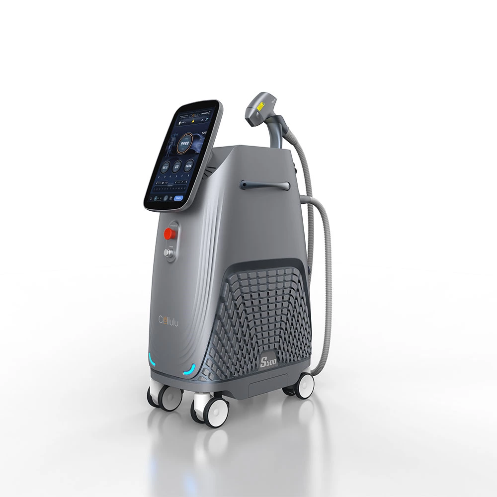
 Ciellulu Laser - Facial Machine Supplier