Seborrheic keratosis

Seborrheic keratosis
Seborrheic keratosis, also known as senile warts, senile plaques, and basal cell papilloma, is the most common benign skin tumor in clinical practice. It is common in middle-aged and elderly people because of benign epidermal hyperplasia caused by hyperplasia of keratinocytes. It is common in the face, back, and back of the hands. It has been reported that the disease has a clear familial tendency, and it is speculated that the disease may be an autosomal dominant genetic disease with incomplete penetrance. Although the disease is common in clinical practice, there are few statistical reports on its gender or racial tendency and regional distribution. The disease is more common in the Caucasian population, with the same incidence in men and women. The disease is rare in people under 40 years old.
1. Etiology and pathogenesis
The disease may be related to sunlight exposure and skin aging.
2. Clinical manifestations
The disease mostly occurs after the age of 40, and is common in the scalp, face, trunk, upper limbs, back of the hands, but does not involve the palms and soles. It starts as a light brown macules or flat papules with a smooth or slightly papilloma-like surface. They increase in size and number with age, with a diameter of 1mm~1cm, or several centimeters, with clear boundaries and a papilloma-like surface. There are greasy scabs on the surface, which are easy to scrape off. Some lesions may be very pigmented, dark brown or black. The color of old lesions varies greatly, and may be normal skin color, light brown, dark brown or black. This disease can be single, but usually multiple, with no subjective symptoms and occasional itching. The skin lesions develop slowly and rarely become malignant.
Clinically, there are several special types:
(I) Irritant seborrheic keratosis
Irritant seborrheic keratosis occurs at the site of cortical overflow or friction. The skin lesions may become irritated, the base becomes red, and the surface is irregularly proliferating.
(II) Eruptive seborrheic keratosis
Eruptive seborrheic keratosis occurs suddenly and increases rapidly in a short period of time. Attention should be paid to whether there are concurrent visceral tumors.
(III) Plaster keratosis
Plaster keratosis mainly occurs in the elderly, and is prone to occur in the lower limbs. The skin lesions are multiple keratotic papules, which are easy to peel off and do not bleed.
3. Pathological characteristics
The basic characteristics of this disease are outward growth, excessive keratinization, thickened acanthosis, papilloma-like hyperplasia, and pseudohorn cysts. Some lesions have many black particles in the proliferating keratinocytes.
4. Diagnosis and differential diagnosis
This disease is not difficult to diagnose based on age, clinical manifestations, etc. Some early lesions resemble flat warts; lesions in exposed areas are easily confused with solar keratosis, lesions with very dark pigments need to be differentiated from melanocytic nevi, and lesions with inflammation or irritation may be similar to basal cell carcinoma, squamous cell carcinoma or malignant melanoma, which can be differentiated by histopathological examination.
5. Treatment
This disease should be treated differently according to the patient's requirements and different skin lesions. For atypical skin lesions that need to be differentiated from melanoma, it is recommended to perform a pathological biopsy after surgical excision. If the patient has cosmetic requirements, laser treatment is feasible.
(I) Q-switched laser
For seborrheic keratotic lesions that are not significantly higher than the skin surface and are mainly characterized by increased pigmentation, Q-switched laser can be used for treatment. Q-switched alexandrite laser (wavelength 755nm), Q-switched ruby laser (wavelength 694nm), and 0-switched frequency-doubled Nd:YAG laser (wavelength 532nm, 1064nm) can all achieve good therapeutic effects.
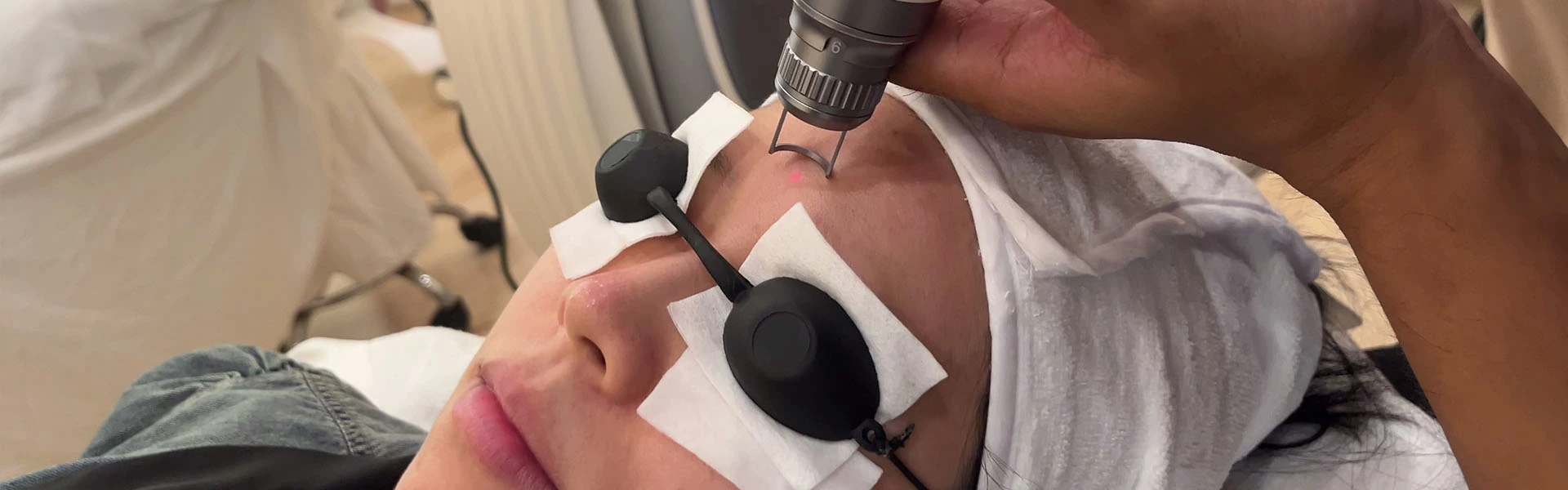
The preoperative preparation for treatment is the same as that for freckles. The treatment dose is generally greater than that for freckles. The scab will fall off 7~14 days after surgery. Pigmentation may occur after surgery. Pay attention to sun protection and it may disappear on its own. If a part is not removed after the first treatment, the second treatment should be performed 6 months later when the pigmentation after laser disappears completely.
(II) CO2 laser treatment
For lesions that are significantly higher than the skin surface, some traditional treatment methods can be used, such as freezing, CO2 laser, etc. However, improper operation may leave scars. If the skin lesions are on the face or the patient has high requirements for beauty, high-energy ultra-pulsed CO laser treatment can be used.
During the treatment, the treatment area is routinely disinfected. Generally, anesthesia is not required. For those who are sensitive to pain, lidocaine cream can be used to seal the surface for anesthesia. Choose a suitable dose. Too low a treatment dose will lead to the formation of a carbonized layer, which will increase the degree of thermal damage and make the depth of treatment difficult to control. During the treatment, the wound surface should be repeatedly wiped with a saline cotton swab to remove the remaining carbonized layer, and the skin layer should be clearly seen before vaporization. Repeat several times until the skin lesions are completely removed, the normal tissue is exposed, and the superficial dermis is reached. Apply antibiotic ointment after surgery. Generally, scabs will form 2 to 3 days after surgery, and scabs will fall off 7 to 10 days after surgery. Sunscreen can be applied after the skin falls off.
Adverse reactions: If the treatment is too deep during Co2 laser treatment, scars will form after healing.
Source: Seborrheic keratosis
 Ciellulu Laser - Facial Machine Supplier