Nevus of Ito

Nevus of Ito
Nevus of Ito was first reported by Ito in 1954, hence the name. Nevus of Ito is a pigmented lesion that occurs in the distribution areas of the posterior supraclavicular nerve and the lateral brachial cutaneous nerve, such as the neck, supraclavicular region and upper arm on one side. The skin in the lesion area is characterized by brown-blue patches or plaques, so it is also called the acromion deltoid brown-blue nevus. This nevus is more common in women than in men, accounting for about 80% of patients. About 60% of patients have lesions at birth.
1. Etiology and pathogenesis
Nevus of Ito is mostly caused by abnormal distribution of melanocytes in the skin.
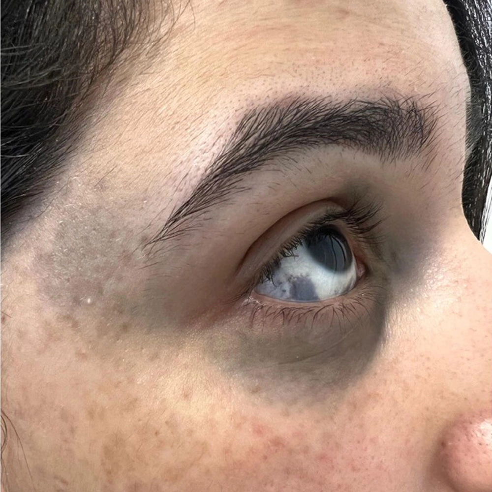
2. Clinical manifestations
Light blue, gray-blue, light brown, dark brown or blue-brown macules or patches occur on the skin of the neck, supraclavicular region and upper arm, and are mottled. Generally, the center of the patch is dark and the edges gradually become lighter, or the center is a patch and the edges are spots, or the entire lesion is a patch of varying density, and the boundaries are often not very clear. Occasionally, some areas of the lesion may be slightly raised or have blue-black protrusions ranging in size from millet to mung beans. Some cases may be accompanied by the same side or bilateral nevus of Ota, or even nevus of Ota and port-wine stain at the same time.
3. Pathological characteristics
The microscopic characteristics of this nevus are the same as those of nevus of Ota. A large number of rhombus-shaped, dendritic and stellate melanocytes are gathered between the collagen fiber bundles in the upper part of the reticular layer of the dermis, which may involve the upper part of the dermis or subcutaneous tissue. Melanocytes can be seen in a few lesions.
4. Diagnosis and differential diagnosis
It is not difficult to diagnose based on clinical manifestations. It must be differentiated from Mongolian spots, blue nevus and Baker's nevus.
(I) Mongolian spots
Mongolian spots are present at birth and can disappear on their own within a few years. The lesions can occur anywhere on the body.
(II) Café au lait spots
Café au lait spots are the skin manifestation of neurofibromatosis. They are multiple and circular pigmented spots on the skin surface with yellow-brown color and varying sizes. Blocks,
(III) Baker's nevus
Ito's nevus skin lesions are blue-brown or dark brown patches, which are more likely to occur on the shoulders, back and upper limbs, with a smooth and hairless surface; Baker's nevus is mostly brown or yellow-brown patches, which are more likely to occur on the back, shoulders and chest, with coarse hair on the surface and slightly thickened skin texture.
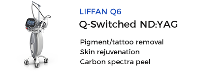
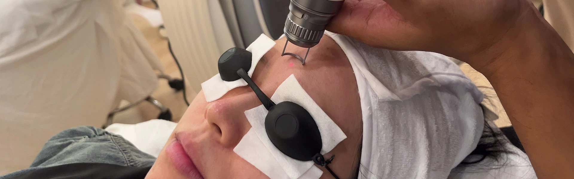
5. Treatment
The treatment is the same as that of Ota's nevus
Source: Nevus of Ito

 Ciellulu Laser - Facial Machine Supplier