Mongolian spots

Mongolian spots
Mongolian spots, also known as birthmarks, are congenital dermal melanocytosis. Because babies are born with them, they are also called moles. Histologically, melanocytes can be seen staying deep in the dermis, so it is also called dermal melanosis. Mongolian spots can occur in any part of the body, but are more common in the lumbar and buttocks. Because melanin particles are located in deeper parts, they appear special gray-green or blue under the Tyndal effect of light. As the baby grows, the color of Mongolian spots gradually fades or disappears, and there is no harm to the body, so no special treatment is required.
1. Etiology and pathogenesis
Mongolian spots are mostly caused by melanocytes staying deep in the dermis during the migration from the neural crest to the epidermis during the embryonic period.
2. Clinical manifestations
Mongoloid spots are usually present in the fetus, deepen after birth, and then gradually fade. Pigmentation spots are almost always limited to the lumbar sacral region and buttocks, occasionally seen on the thigh side or even the shoulder, and are gray-green, blue or blue-black, round, oval or irregular in shape, with unclear edges, and a diameter ranging from only a few millimeters to more than ten centimeters. They are mostly single, and occasionally multiple. Except for the change in pigmentation, the affected area has no abnormalities, and the skin texture is also normal. It usually disappears on its own without leaving any traces at the age of 5 to 7 years old, and occasionally persists in adulthood or even expands. Mongolian spots are common in Orientals or blacks, and the incidence in Mongolian infants can reach more than 90%. It is rare in people of other races.
3. Pathological characteristics
Histopathological examination shows that the dermis, especially the lower half, is full of melanin granules of melanocytes, whose dendrites are significantly elongated and thinned, often in a microwave shape, roughly parallel to the skin surface, and widely scattered between the collagen fiber bundles. The melanin granules contained are positive for DOPA reaction, indicating that they are not melanocytes in the dermis. Under the electron microscope, it can be seen that these melanocytes contain countless completely melanized melanosomes.
4. Diagnosis and differential diagnosis
Based on the characteristic skin lesions and specific disease progression after birth, it is generally not difficult to diagnose. Differential diagnosis is required from blue nevus and nevus of Ota.
(I) Blue nevus
Blue nevus is generally darker in color, with fairly clear borders and small dome-shaped nodules; cellular blue nevus is a large nodule or plaque, all slightly raised on the skin surface. Mongolian spots are present at birth or may begin in early childhood, and may be accompanied by compound nevus or malignant transformation.
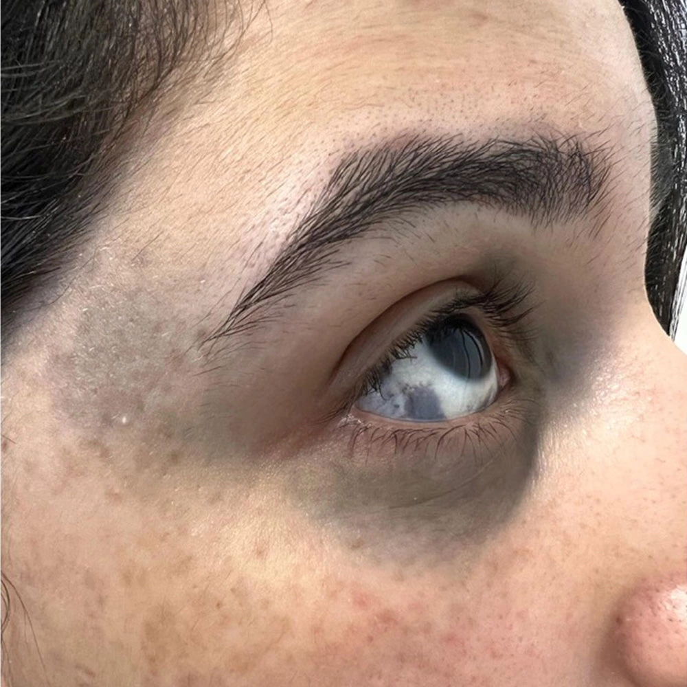
(II) Nevus of Ota
Nevus of Ota often grows on the face, and the lesions are often mottled, mixed with brown and blue spots.
5. Treatment
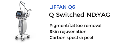
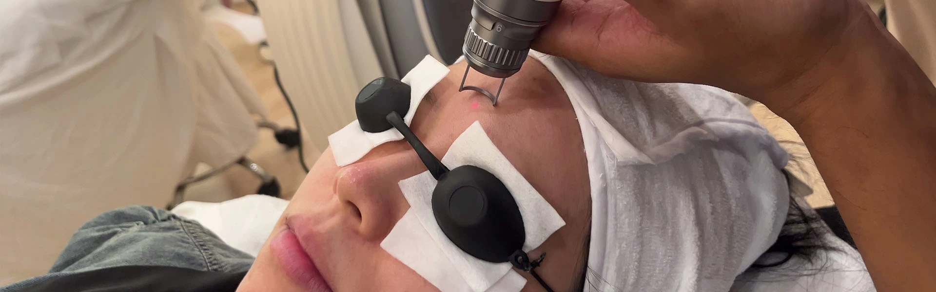
Generally, it can disappear on its own and no treatment is required. If it does not disappear, Q-light laser treatment can be used, and it can be cured after several treatments. The treatment is the same as that of nevus of Ota.
Source: Mongolian spots

 Ciellulu Laser - Facial Machine Supplier