Chloasma

Chloasma
Chloasma is a pigmentation spot that occurs frequently on the cheeks and forehead of women of childbearing age. It is generally symmetrically distributed and can exist for a long time or develop chronically. In traditional Chinese medicine, it is also called "liver spots" and "black spots". Those caused by pregnancy are also called pregnancy spots, which can disappear after delivery.
1. Causes and pathogenesis
The causes and pathogenesis of chloasma are not yet fully understood. Current studies have shown that ultraviolet radiation, cosmetics, Chapter 2 Laser treatment of pigmented skin diseases
Pregnancy, endocrine disorders, certain chronic diseases, certain drugs, insomnia, long-term negative emotions and transmission are all related to the onset of chloasma.
Studies have shown that the secretion of melanocyte-stimulating hormone (MSH) increases during pregnancy, which can lead to active melanocyte function. It has been proven that estrogen can stimulate melanocytes to secrete melanin granules, and progesterone can promote the transport and diffusion of photophores. Pregnancy spots are caused by the combined action of these two hormones. Women who take oral contraceptives and some chronic gynecological diseases such as menstrual disorders, dysmenorrhea, adnexitis, infertility, etc. can also induce chloasma, and it is believed that its occurrence is related to abnormal estrogen and progesterone in the body.
Ultraviolet rays are also an important triggering factor of chloasma. Ultraviolet rays can increase the activity of tyrosinase, stimulate the division of melanocytes, and proliferate melanocytes in the irradiated area. Therefore, this disease is often induced or aggravated after sun exposure in summer. It also often occurs in patients with some chronic diseases such as liver disease, chronic alcoholism, thyroid disease, and visceral tumors, indicating that this disease is related to endocrine factors such as ovaries, pituitary glands, and thyroid glands. In addition, oral drugs such as chlorpromazine and phenytoin sodium can also induce chloasma. Trace elements copper and zinc also have a certain effect on the onset of chloasma. In the past, it was believed that the onset of chloasma was mainly attributed to melanin metabolism disorders. Recently, many scholars have shown that in addition to pigment metabolism disorders, impaired skin barrier function in the affected area, inflammatory reactions, and local microvascular dysfunction all play an important role in the onset of chloasma.
2. Clinical manifestations
Chloasma is more common in women, especially those of childbearing age, but can also occur in men. The lesions are often symmetrically distributed on the zygomatic and cheek areas and are butterfly-shaped, commonly known as "butterfly spots". They can also affect the forehead, nasal dorsum, periorbital or chin areas. The color is light yellow-brown, dark brown or dark coffee-colored with irregular shapes and clear or diffuse edges. The lesions generally do not affect the eyelids and oral mucosa, and there are no subjective symptoms. The color of the lesions often deepens after exposure to ultraviolet rays, and they often worsen in spring and summer and alleviate in autumn and winter. In addition, factors such as mood, sleep and endocrine changes can also cause slight changes in the color of the lesions. The course of the disease is uncertain and can last for months or years.
1. Traditional classification Chloasma is divided into three types based on the distribution of lesions.
(1) Midface type: lesions are distributed on the forehead, cheeks, upper lip and nose.
(2) Cheek type: lesions are mainly located on the cheeks and nose. (3) Mandibular type: The lesions are mainly located on the mandible, occasionally affecting the "V" area of the neck.
2. Foreign classification Foreign scholars divide this disease into four types based on the color changes observed by the Wood's lamp and the distribution of melanosomes.
(1) Epidermal type: The pathological changes are mainly manifested as pigmentation in the basal layer, spinous layer, granular layer and even stratum corneum of the epidermis. The dendritic processes of melanocytes extend upward above the basal layer. The color contrast between the lesion area and the non-lesion area increases when observed by the Wood's lamp.
(2) Dermal type: The pathological changes are manifested as melanophages around the blood vessels in the dermis. The color contrast between the lesion area and the non-lesion area does not change when observed by the Wood's lamp.
(3) Mixed type: The color contrast of some lesions increases when observed by the Wood's lamp, while some lesions do not change.
(4) No change type: Obvious lesions can be observed under visible light, but there is no increase in pigmentation under the Wood's lamp. The histological manifestation is pigmentation in the dermis.
3. Pathological characteristics
The melanin formation in the basal layer and spinous layer of the epidermis of this disease is active, and melanin increases, but there is no melanocyte proliferation; free melanin particles can be seen in the upper dermis or are phagocytosed by melanocytes, and there is no inflammatory cell infiltration.
4. Diagnosis and differential diagnosis
This disease is more common in young and middle-aged women. The skin lesions mainly occur on the face, mainly in the zygomatic region, cheeks, and cervix. It has the characteristics of yellow-brown skin lesions and worsening in summer. It is generally easy to diagnose. This disease is mainly differentiated from the following diseases. 1. Freckles are small, scattered and not fused. There is often a family history. They often occur in childhood and are more common in adolescent women. They are obvious in summer and fade or disappear in winter.
2. Reil's melanosis It often occurs on the forehead, zygomatic region and neck side of the face. There are gray-purple to purple-brown dot-like spots,
with powdery small scales attached, which can later merge into pieces. There may be inflammation in the early stage. 3. Brown-blue nevus of the zygomatic region The skin lesions are brown-blue spots with a diameter of 1~5mm. The spots do not merge into a piece. They usually appear in women around 20 years old.
4. Ota nevus It usually occurs at birth or in childhood. The skin lesions are unilaterally distributed and may involve the eyeball and mucous membranes. They are light blue, gray-blue or blue-black patches.
5. Treatment
Due to the complexity of the occurrence of melasma, there is no broad consensus on the treatment of melasma. The following are the common treatment methods.
(I) General treatment
First of all, the factors that induce the disease should be eliminated as much as possible: sun exposure, contraceptives, stress, insomnia or other related chronic diseases. Protect the facial skin, avoid excessive cleaning and excessive stimulation, pay attention to moisturizing and sun protection, etc.
(II) Drug treatment
1. Western medicine treatment, mainly vitamin C, vitamin E, tranexamic acid, glutathione, etc.; for vascular or vascular dominant melasma, anti-inflammatory drugs and drugs that improve vascular function are also needed.
2. Treatment with traditional Chinese medicine: Generally, it is necessary to differentiate and classify the drugs, such as Xiaoyaosan for liver depression and qi stagnation, Shenlingbaizhusan for spleen deficiency and dampness accumulation, Liuweidihuangwan for liver and kidney yin deficiency, Taohongsiwutang for qi stagnation and blood stasis. 3. Local medication: For non-vascular chloasma lesions, a certain concentration of hydroquinone, azelaic acid, vitamin A acid and other depigmenting agents can be given locally, but it should be noted that these drugs may cause local skin irritation, post-inflammatory pigmentation or uneven pigmentation and other side effects. 20%~50% concentration of fruit acid peeling treatment can also be used for non-vascular dominant chloasma; local induction treatments such as arbutin or L-vitamin C can also have the effect of lightening chloasma.
(III) Laser treatment
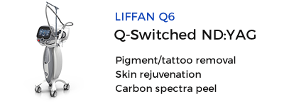
The use of laser for the treatment of chloasma has always been controversial. Because some patients with chloasma will have obvious post-inflammatory pigmentation reaction after laser treatment, or short-term recurrence may occur after treatment. As the research deepens, some domestic experts divide chloasma into vascular type and pigment type, and our choice of the timing of laser treatment for chloasma has gradually become clear. Pigmented chloasma is the consequence of inflammation, while vascular type is a sign that inflammation is occurring. When inflammation is occurring, the use of laser treatment will lead to aggravation of inflammation, which in turn makes melanocytes more active and produces post-inflammatory pigmentation. Therefore, we only advocate the use of laser treatment when chloasma is in the pigment stage. 1. Q-switched laser The wavelengths are 1064nm, 755nm, 694nm and 532nm, and the pulse width is 5~10ns. The principle of removing chloasma pigment particles is still based on selective photothermal effect, that is, pigment particles selectively absorb light of a certain wavelength and then rapidly expand and rupture to form small fragments, which are then engulfed by phagocytes in the body and excreted from the body, while normal tissues will not be ruptured or damaged.
Since melasma is characterized by abnormally active pigment cells in pathophysiology, some scholars have proposed a new theory of laser treatment for melasma based on the experience gained from previous laser exploration of melasma treatment: subcellular selective photothermolysis. Since melasma pigment cells are active, in order to minimize the damage of laser to normal skin tissue and basement membrane, thereby avoiding the aggravation of melasma, the energy selection is to selectively photoblast only the pigment particles in the pigment cells, and try to avoid or reduce the activation of pigment cells. The pigment cell function is inactivated or inhibited through multiple photoblasts of small doses. At the same time, multiple photoblasts of pigment particles can make the pigment particles smaller and more conducive to phagocytosis and excretion.
Q-switched 532nm, 694nm and other short-wavelength lasers are more absorbed by epidermal melanin, causing tissue cell damage. During the repair process, pigmentation is easily caused due to active reactive melanin production. In the past, Q-switched 1064nm and 755nm light treatments for melasma used small spots, high energy, and few treatments, which usually caused damage to the surrounding skin tissue and basement membrane, and were also prone to the side effect of increased pigmentation. However, in recent years, Q-switched emerald 755nm lasers and Q-switched Nd:YAG1064nm have used large spots, low energy, and multiple treatments, which have achieved good results, with mild tissue reactions, avoiding and reducing post-inflammatory pigmentation, and gradually becoming the mainstream laser treatment options for Asians.
The treatment steps are as follows:
(1) Preoperative precautions.
① Before surgery, the physician must first determine that the melasma lesions are in a stable pigmentation stage.
② No history of sun exposure within 1 month before treatment.
③ For those with facial skin inflammation, the facial inflammation should be controlled first.
(2) Clean the face before surgery. Chloasma patients should use a mild facial cleanser to clean their faces before surgery, and disinfect the surgical area with routine Sanisol.
(3) Surface anesthesia/general anesthesia. No anesthesia is required.
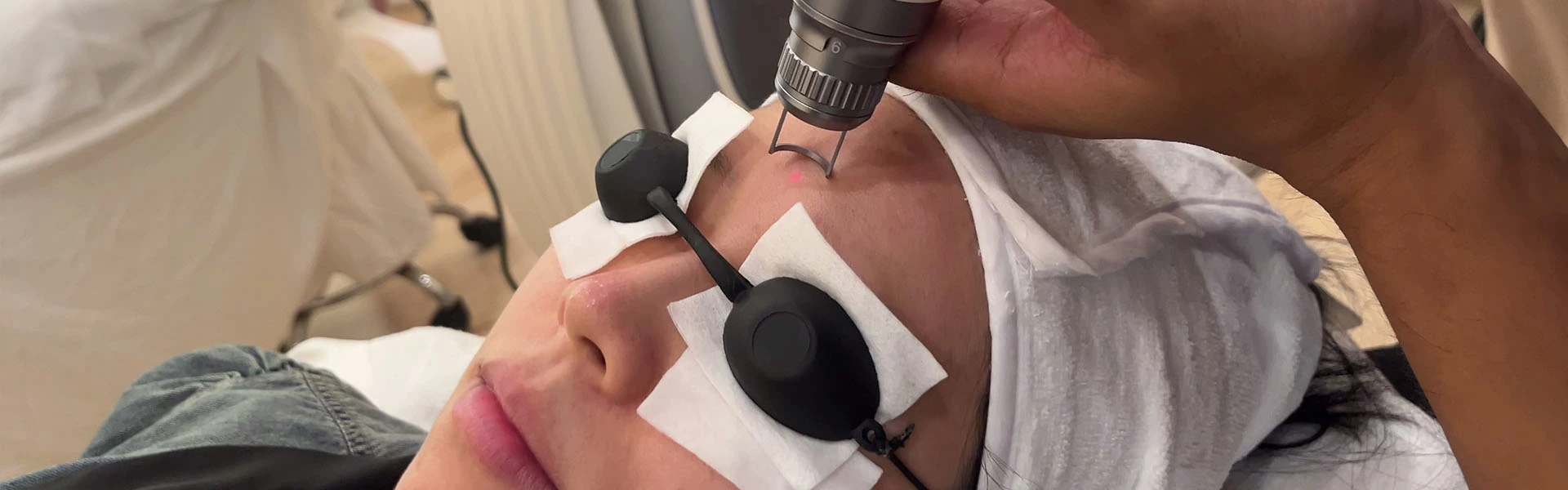
(4) Eye protection. The operator and the patient should wear special goggles.
(5) Intraoperative treatment response. Generally, a 6~8mm spot is used, and the energy density is generally 2~3mJ/cm?. After irradiation, the skin will show mild flushing.
(6) Treatment of the surgical area after surgery. Immediately after surgery, a medical skin care mask or induction treatment with facial skin barrier function repairing function is given to prevent micro-injury symptoms such as skin dryness and sensitivity caused by laser.
(7) Postoperative precautions.
① After laser treatment, the face should be washed and makeup should be applied in a gentle and friction-free manner, and facial skin should be moisturized and repaired under the guidance of a doctor.
②) During the interval between two treatments, sunscreen should be strictly used to prevent sun exposure.
③ The incidence of side effects of this laser treatment method is low. Occasionally, pigmentation aggravation or secondary hypopigmentation spots are generally recovered in 2~6 months. If pigmentation occurs, the next treatment should be chosen after the pigmentation is restored. (4) The treatment frequency is generally once a week, and most of the time, 5~10 treatments are required.
2. Fractional laser treatment Fractional laser technology is a technical concept between invasive ablative and non-invasive non-ablative treatment methods. Fractional lasers are divided into two categories: non-ablative fractional lasers and ablative fractional lasers. Its mechanism of action is an extension of the traditional selective photothermal theory, that is, fractional photothermal effect. Tissue water is the target color base of the fractional laser. The fractional laser generates a small beam arranged in an array to act on the skin. After the skin tissue water absorbs the laser energy, it forms multiple micro-columnar thermal damage zones (micro-thermal damage zones MTZ), which then cause a series of skin biochemical reactions, achieving the effects of skin tightening, skin rejuvenation and spot removal. The MTZ has a diameter of 50~150pm and a depth of 400~1000um. The MZ generated per square centimeter of skin in the treated area may be different due to the different beam dot designs of the instrument. For example, the FraxeSR series has two densities of 125MTZ/cm' or 250MTZ/cm'. Only 12%~20% of the skin in the treated area forms MTZ. The dot laser forms a circular tissue coagulation zone or thermal damage zone around each MTZ, and the periphery is undamaged normal tissue, which can accelerate the repair and regeneration of the damaged skin, shorten the wound repair period after treatment, and reduce pigmentation.
The dot laser used in the treatment of melasma is a non-ablative dot laser with a wavelength of usually 1540nm or 1550nm. Unlike ablative laser, it does not damage the epidermal stratum corneum, and the remaining epidermal tissue coagulates but does not vaporize. Its MTZ includes the epidermal tissue under the stratum corneum and the dermal tissue at different depths. This not only retains the barrier function of the skin, but also directly damages the melanocytes, melanin granules and keratinocytes of melasma lesions through the micro-heat zone generated by the dot laser. This is a relatively safe and effective option for treating melasma. It is currently approved by the FDA for the treatment of melasma. However, fractional laser treatment also has the problem of pigmentation and recurrence. The treatment operation steps of fractional laser are as follows: (1) Preoperative precautions and operation: Same as Q-switched laser treatment of melasma. (2) Intraoperative treatment response. The specific parameter settings during treatment should be adjusted by the clinician according to the patient's skin type, melasma lesions and response to treatment. In principle, it is recommended to choose a small spot, slightly lower energy and low dot density. Generally, the treatment endpoint is the appearance of slight redness and edema in the lesion area. (3) Postoperative treatment of the surgical area. Immediately after surgery, apply an ice pack to the treatment area for 15 to 30 minutes. Apply skin repair products such as epidermal growth factor.
(4) Postoperative precautions.
① Avoid washing your face or bathing 2 to 3 days after laser surgery.
② The surgical area may peel or have slight scabs. Generally, the scabs will fall off after 7 to 10 days. Be careful to let them fall off naturally. After the scabs fall off normally, you can use skin care and makeup normally. During the treatment, cooperate with facial hydration care to achieve the best effect after treatment.
③ During the interval between two treatments, you need to strictly use sunscreen to prevent sun exposure.
It is recommended that there is a 1-month interval between two treatments. Generally, after 4 treatments, the efficacy should be evaluated and whether to continue this treatment method should be evaluated.
Some patients will still have temporary pigmentation, which often takes 2 to 6 months to recover. Suspend laser treatment during the recovery period of pigmentation.
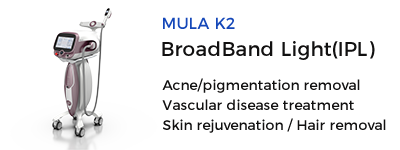
3. Intense pulsed light therapy (IPL) Intense pulsed light (IPL) is an incoherent wide-spectrum visible light emitted by a xenon lamp with a wavelength of 500~1200nm, select the corresponding filter according to the patient's skin quality and skin lesions, screen out light of different wavelengths for the treatment of skin lesions, the pulse width is adjustable, and 1~3 pulses can be selected for each firing. Its working principle is the same as that of Q-switched laser, which is still selective photothermal effect, so pigmentation problems are also its indications. The IPL pulse width is a millisecond-level light source, which cannot instantly concentrate the peak energy to blast melanosomes, and cannot effectively destroy the melanin particles in the dermis. Compared with Q-switched lasers, IPL has a long pulse width and low energy, causing less tissue damage reaction and less pigmentation after treatment. In recent years, large-sample observations of IPL treatment of melasma have been reported in China, and it is believed that IPL is safe and effective for the treatment of refractory melasma in Asians. At present, the fourth-generation IPL uses optimized pulse technology (O0PT), with uniform pulse energy control, flat waveform top, no energy peak and energy decay. The treatment is mild, safe and effective. Yanmshita observed that melanocytes are not destroyed after IPI irradiation and can quickly recover their activity. He believes that PL can temporarily remove epidermal spots, but to maintain the therapeutic effect, drugs or effective laser treatment should be added to inhibit the activity of melanocytes. The precautions before and after IPL treatment and the treatment endpoints are roughly the same as those of 0-switching laser. After IPL irradiation of melasma lesions, the color of the spots may immediately deepen. This is because after IPL irradiates the lesions, the melanin particles in the basal layer of the epidermis quickly move up to the surface of the skin, and necrotic fragments of cells with aggregated melanin particles appear. The erythema and pain during and after IPL treatment are mild and generally disappear within 1 day. Most patients have slight scabs, which generally fall off within 1 to 2 weeks. Cosmetics can be used immediately after treatment without wound care, and there is no infection or scar formation. After inflammation after IPL treatment Pigmentation is lighter than Q-switched laser and ablative laser.
Source: Chloasma
 Ciellulu Laser - Facial Machine Supplier