Cheek Acquired Dermal Melanocytosis
Cheek Acquired Dermal Melanocytosis
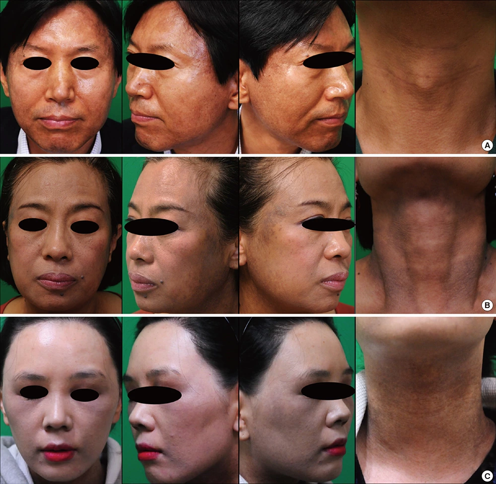
Fig. 1. Clinical photographs. Pigmentation was prominent on lateral face, perioral and neck. (A) M/51 Brownish mottled patch developed on face and neck 6 months ago. (B) F/55 Slate-greyish colored patch developed for 12 months on lateral face and neck. (C) F/33 Grey colored patch slowly developed over 5 years.
Cheek Acquired Dermal Melanocytosis is also known as Acquired Nevus of Ota, Acquired Bilateral Nevus of Ota-like Macules, Acquired Facial Dermal Melanocytosis, and Acquired Localized Facial Dermal Melanocytosis. It was first reported by Ho in 1984 and is also called Hoi Nevus. The main feature is the symmetrical distribution of black-grey macular pigmentation on the cheek. It was previously considered a variant of Nevus of Ota, but is now thought to be a separate disease. This condition is more common in women, with a reported male-to-female ratio of 1:12.8-17.7. The average onset age is 30 years (ranging from 6 to 54 years), with 89% of cases being Fitzpatrick skin type IV, and the remainder being types II and III.
I. Etiology and Pathogenesis
The exact cause and mechanism of this disease are currently unclear. It is speculated that during embryonic development, melanocytes migrating from the neural crest to the epidermis may fail to pass the dermo-epidermal junction and remain in the dermis. These immature dermal melanocytes can later be activated by ultraviolet (UV) radiation, radiation, hormones, and chemical factors, leading to the formation of the lesions. Keratinocytes, when exposed to UV radiation, release endothelin-1 and granulocyte-macrophage colony-stimulating factor, which can accelerate melanogenesis. Mizushima also believes that immature melanocytes can become activated later even without any stimulation. Studies indicate that genetic susceptibility and environmental risk factors (such as the use of harmful cosmetics and UV exposure) also play important roles in the pathogenesis of this disease. 20.9% to 25% of patients have a family history of the condition. Domestic scholars, including He Li, have shown that dermal melanocytes in CADM patients express androgen receptors, although serum androgen levels are normal.
II. Clinical Presentation
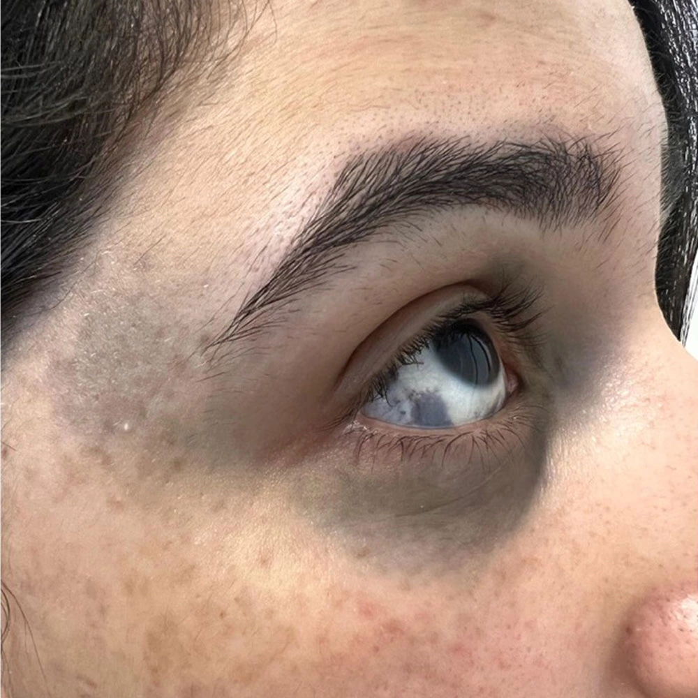
This disease is acquired and affects the face, predominantly the zygomatic region near the outer lower eyelid. Occasionally, it can appear on the eyelids, nasal wings, and forehead. The lesions are grey-brown, black-grey, or blue-brown pigmented macules, 1-5mm in diameter, round, oval, or irregular in shape, with well-defined borders. The number of lesions varies from a few to several dozen, averaging between 10 and 20, and they are usually bilaterally symmetrical. The eyes and oral mucosa are not affected, and the condition often coexists with melasma. Patients do not experience any subjective symptoms.
III. Histopathological Characteristics
The epidermis appears normal, with primary changes observed in the upper dermis, particularly the lower papillary layer. Scattered small, spindle-shaped melanocytes aligned parallel to collagen fibers are present, and they are positive for dopa staining. The dermal structure is otherwise normal. Electron microscopy reveals numerous melanosomes of varying sizes at different stages of development within melanocytes.
IV. Diagnosis and Differential Diagnosis
Diagnosis is generally based on clinical presentation but needs to be differentiated from the following conditions:
1. Nevus of Ota Nevus of Ota typically presents with unilateral distribution along the ophthalmic and maxillary branches of the trigeminal nerve, with early onset, usually at birth or before age 2. The lesions are confluent pigmented macules often accompanied by eye and oral mucosal involvement. Histopathological differences include more melanocytes dispersed throughout the dermis without alignment with collagen fibers, unlike in CADM.
2. Freckles (Ephelides) Freckles appear earlier in life, are smaller, lighter in color (yellow-brown), and exhibit a seasonal variation, becoming more pronounced in summer. Histopathologically, freckles show increased melanin in the basal layer of the epidermis without an increase in the number of melanocytes.
3. Melasma Melasma predominantly affects the cheeks, zygomatic region, forehead, and upper lip, with lesions merging into larger patches of light to dark brown color, which intensify during summer. Unlike CADM, melasma does not involve the eyelids and has a different histopathological profile.
V. Treatment
Given the abnormal pigment distribution in the dermis, Q-switched laser treatment is preferred:
1. Q-Switched Ruby Laser (694nm) Wavelength 694nm, pulse width 20-40ns.
Laser Treatment Steps:
Pre-Treatment Care:
- If the lesion area has concurrent sunspots or melasma, treat these first to reduce the risk of post-inflammatory hyperpigmentation and prevent exacerbation by laser stimulation.
- Control any facial inflammation to reduce the likelihood of post-treatment hyperpigmentation.
Procedure:
- Cleanse the face similarly to the preparation for Nevus of Ota treatment.
- Apply surface or general anesthesia if needed. For small lesions, anesthesia may not be necessary, but for larger or more sensitive areas, topical or local anesthesia can be used.
- Protect the eyes with special goggles or corneal shields during treatment.
- Use an energy density of 5-8J/cm², slightly overlapping laser spots, and treating all pigmented areas without missing any lighter or smaller spots.
Post-Treatment Care:
- Manage post-treatment inflammation and provide patient education to prevent pigmentation changes.
- Monitor for post-inflammatory hyperpigmentation, which may take six months or longer to resolve. Advise patients on the importance of patience and adherence to follow-up treatments.
2. Q-Switched Alexandrite Laser (755nm) The Alexandrite laser at 755nm causes minimal pain and usually does not require anesthesia. Treatment sites may exhibit mild swelling that subsides within 24 hours. Early treatment yields better results due to better absorption and fewer pigment granules in younger patients.
Early intervention and appropriate laser treatment are critical in managing CADM effectively. The condition, while benign, can have significant cosmetic implications, especially for affected individuals. A thorough understanding of the etiology, clinical presentation, and treatment options ensures optimal patient outcomes.
 Ciellulu Laser - Facial Machine Supplier